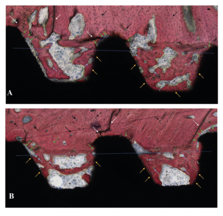
Figure 3.
Histology sections of (A) D1 and (B) D2 at 3 weeks. The blue line represents the theoretic position of the osteotomy outer diameter resulting in the formation of healing chambers between the implant inner diameter, the implant threads, and the osteotomy walls. Interfacial remodeling, depicted by green arrows, and bone microcracking, depicted with white arrows, can be observed at the tip of the implant threads where the initial mechanical interlocking took place upon implant insertion. Woven bone formation is observed to occur within the healing chambers from the surgically prepared native bone, from the implant surface (yellow arrows), and from the central region of the chambers, where bone remodeling sites can be observed (clear blue arrows). In the native bone, cortical shell osteonic structures are depicted with black arrows.

