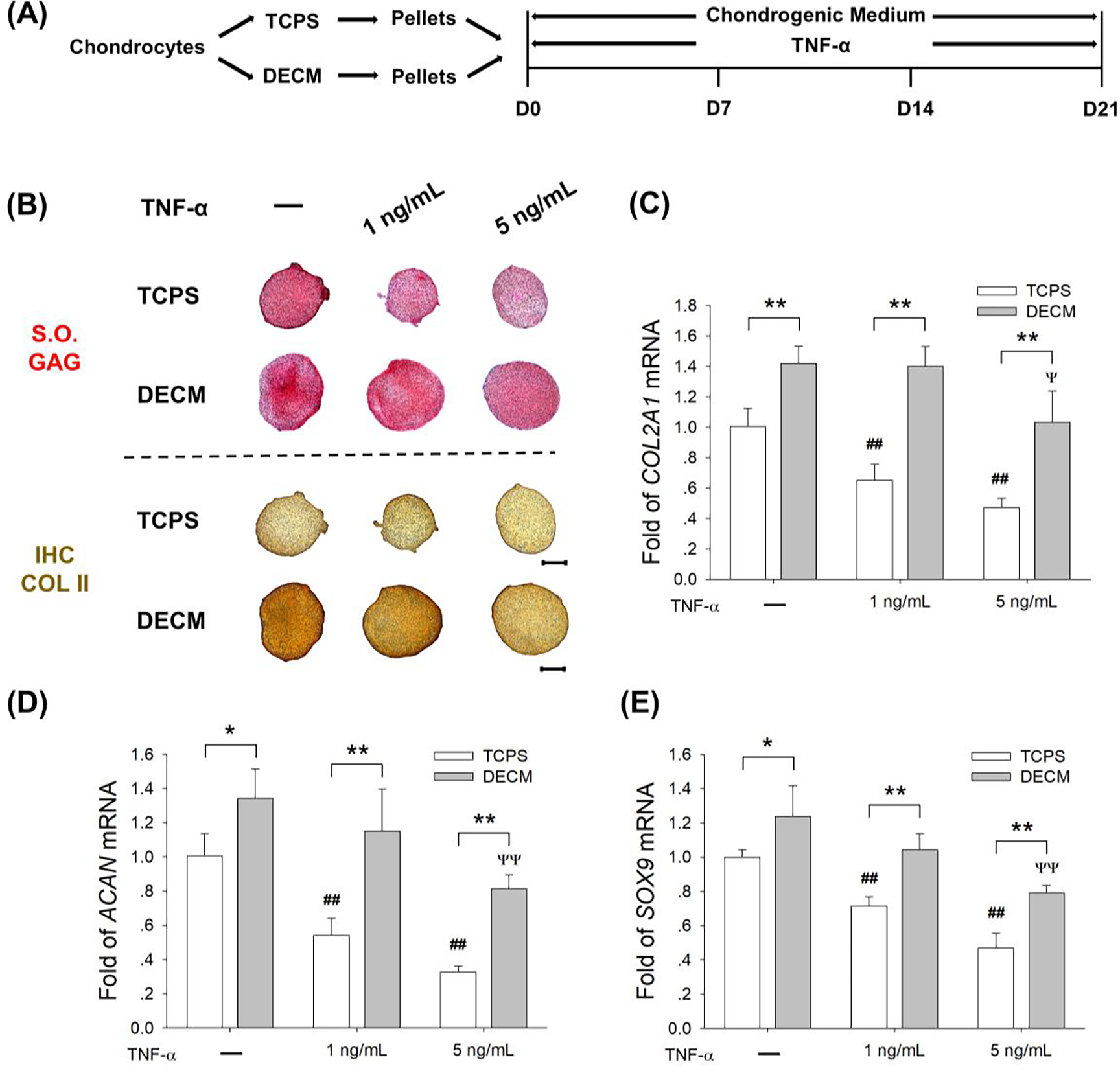
Fig. 4.

DECM expansion of chondrocytes improved their resistance to TNF-α-induced inflammatory stress. (A) TCPS- or DECM-expanded chondrocytes were induced to redifferentiation in the presence of 1 ng/mL or 5 ng/mL of TNF-α for 21 days. (B) Safranin O (S.O.) was used to stain sulfated GAGs and immunohistochemistry staining (IHC) was used to detect COL II. Scale bar = 500 μm. (C-E) The mRNA levels of COL2A1 (C), ACAN (D), and SOX9 (E) were evaluated by RT-qPCR. Values are presented as the mean ± S.E.M. of four independent experiments (n = 4) in RT-qPCR experiments. Statistically significant differences are indicated by * p < 0.05 or ** p < 0.01 between the indicated groups; # p < 0.05 or ## p < 0.01 versus TCPS-expanded pellets without TNF-α treatment; Ψ p < 0.05 or ΨΨ p < 0.01 versus DECM-expanded pellets without TNF-α treatment.
