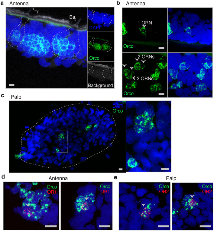
Figure 3.
Expression of SameOrco, SameOR1, and SameOR2 in antenna and palp tissue sections, respectively. RNAscope in situ hybridization was used to characterize the spatial distribution of these putative ORs. (a–c) Probes against SameOrco (green) in antenna sections (a, b) and palp sections (c). Orco+ cells in trichoid (Tr) and basiconic (Ba) sensilla are highlighted by arrows in panel a. Trichoid sensilla containing one, two, and three ORNs are depicted by arrows in panel (b). (d, e) SameOrco probe with the probe against the second most expressed putative SameOR in each tissue type were used. SameOR1 (antenna, panel d) and SameOR2 (palp, panel e) are present only in Orco+ cells. Blue label in all images:DAPI. Scale bars: 10 μm in panels a–c; 15 μm in panels d–e.

