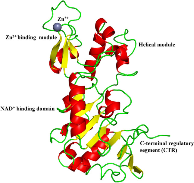
Fig. 1.

Structure of SIRT1 (PDB ID: 4IG9). The NAD+ binding region is made up of a core six-stranded parallel β sheet with strands β1-3 and β7-9, as well as eight helices namely, αA, αB, αG, αH, and αJ-M, that pack against the β sheet core. The helical module is made up of four α helices namely, αC-F, while the Zn2+binding unit is made up of three β strands, β4 to β6 and one helix αI. The CTR creates a β hairpin shape that covers a fundamentally unchanging, hydrophobic region, complementing the β sheet of the NAD+ binding domain
