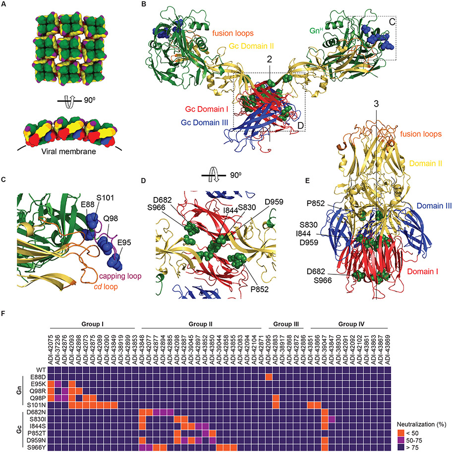
Fig. 4: Epitope mapping of nAbs by escape mutant analysis.
(A) Schematic representation of the organization of lattices of Gn/Gc tetramers in viral particles (top, en face view; bottom, side view). Gn, green; Gn capping loop, purple; Gc domain I, red; Gc domain II, yellow; Gc domain III, blue; Gc fusion loops, orange. The approximate position of the viral membrane is indicated with a black line. (B) Ribbon representation of the ANDV GnH/Gc pre-fusion dimer (modeled from its monomer, PDB: 6Y5W; (14)) with the two-fold molecular axis of the dimeric spike indicated (“2”). Residues exchanged in the neutralization-escape mutants are shown as blue (Gn) or green (Gc) spheres. (C–D) The GnH:Gc and Gc:Gc interfaces in panel B (dotted rectangles) were enlarged for clarity and rotated as indicated to show neutralization-escape mutants in the Gn capping loop (C) and Gc (D). In panel C, the cd fusion loop in Gc associated with the capping loop in Gn is shown in orange. (E) The PUUV Gc post-fusion trimer with the three-fold molecular axis indicated (“3”) (PDB: 5J81; (20)). Labeling of Gc domains and escape mutations as in panel B. (F) Heatmap showing the capacity of mAbs to neutralize rVSV-PUUV-Gn/Gc bearing neutralization-escape Gn/Gc mutants (also see Fig. S5). mAbs were grouped according to competition Groups I-IV (see Fig 3). Group I = ADI-42898 competitors; Group II = ADI-42098 competitors; Group III = ADI-42898/ -42098 non-competitors; Group IV = undefined/ GnH/Gc non-binders. Neutralization averages (n=6) from two experiments.

