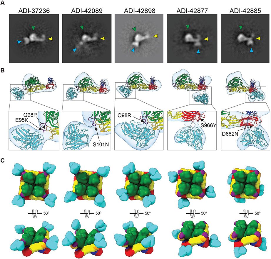
Fig. 5: Negative-stain electron microscopy (nsEM) of scFv:PUUV GnH/Gc complexes.
(A) Exemplary nsEM 2D-classes of PUUV GnH/Gc bound to scFvs of the indicated mAbs. scFvs, turquoise; GnH, green with the capping loop in purple; Gc domain I, red; domain II, yellow; domain III, blue; fusion loops, orange. (B) 3D reconstructions of scFv:GnH/Gc complexes are shown in transparent surface representations (light gray) with the structure of ANDV GnH/Gc docked into the density and pseudo-colored as described in panel A. Corresponding neutralization-escape mutations are shown in the closeups. (C) Modeled interactions of the indicated scFvs with tetrameric PUUV GnH/Gc complexes are shown in surface-shaded representation and colored as in panel A. En face and side views are shown.

