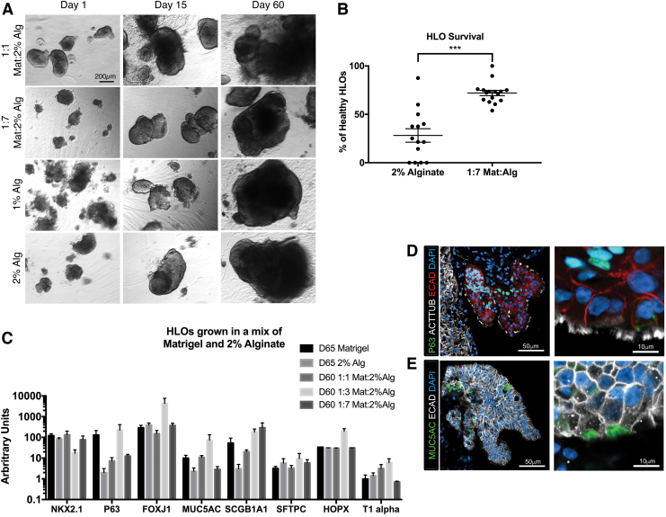
FIG. 3.
Healthy HLOs increased when matrix was added to the alginate. (A) Wholemount images of HLOs cultured in 1:1 Matrigel:2% alginate, 1:7 Matrigel:2% alginate, 1% alginate, and 2% alginate over 60 days. Scale bar applies to all images. (B) HLOs cultured in 1:7 Matrigel:2% alginate had significantly higher initial rate of healthy organoids (72.1% ± 2.7%, n = 16 alginate droplets) compared with 2% alginate (28.2% ± 6.5%, n = 14 alginate droplets). HLOs were quantified after 5 days in the alginate droplet. Error bars represent SEM. (C) HLOs grown in 100% Matrigel, 2% alginate. 1:1 Matrigel:2% alginate, and 1:7 Matrigel:2% alginate for 60–65 days had similar expression of lung marker NKX2.1, airway markers (P63, FOXJ1, MUC5AC, SCGB1A1), and alveolar markers (SFTPC, HOPX, T1alpha). All error bars represent SEM from n = 3 for each group. Data presented as fold change from hPSC control. (D) 1:7 Matrigel:2% alginate HLOs cultured for 60 days had multiciliated cells labeled by ACTTUB (white) covering the epithelial structures labeled by ECAD (red). The P63+ (green) basal cells labeled were on the basal side of the epithelium, forming a pseudostratified epithelium. (E) 1:7 Matrigel:2% alginate HLOs cultured for 60 days had MUC5AC+ (green) mucus producing goblet cells within the epithelium labeled by ECAD (white). Color images are available online.

