FIG. 5.
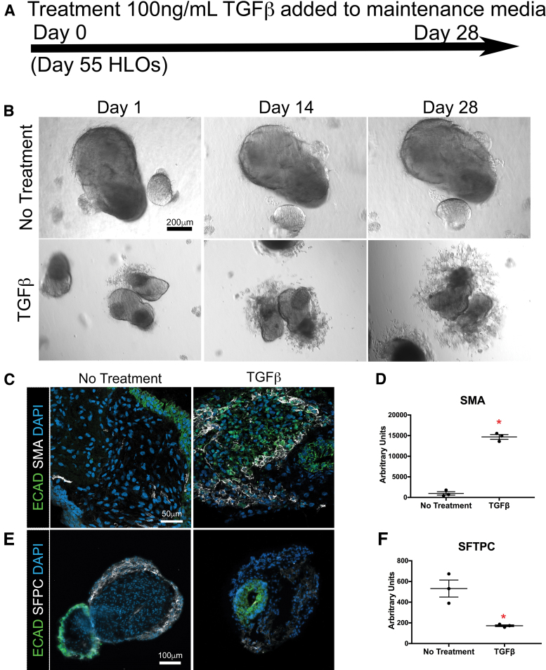
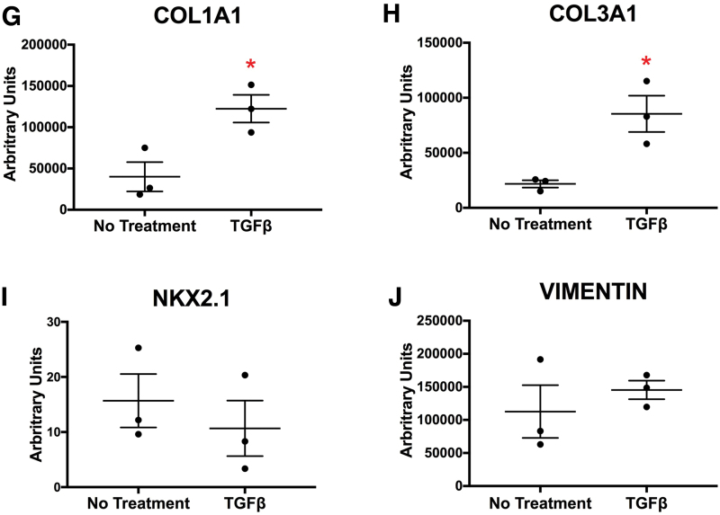
HLOs grown in alginate gained a fibrotic phenotype when treated with TGFβ. (A) Timeline of TGFβ treatment. Treatment started with HLOs cultured for 55 days, then 100 ng/mL of TGFβ was added for 4 weeks. HLOs were cultured for a total of 83 days. (B) HLOs treated with TGFβ had mesenchymal outgrowths compared with the untreated group. (C) There was an increase of SMA+ (white) cells, which label myofibroblasts, in the TGFβ treated group compared with the untreated group. Epithelium was labeled by ECAD (green). (D) There was a significant increase of SMA transcript in the TGFβ-treated group compared with the untreated group. (E) The TGFβ treated group had fewer SFTPC+ (white) cells compared with the untreated group. SFTPC labels type II alveolar cells and alveolar progenitors. Epithelium was labeled by ECAD (green). (F) There was a significant decrease of SFTPC transcript compared with the untreated group. (G, H) COL1A1 and COL3A1 mRNA expression were significantly higher in the TGFβ-treated group compared with the untreated group. (I, J) TGFβ-treated and untreated groups had similar mRNA expression of early lung marker NKX2.1 and mesenchymal marker vimentin. All error bars represent SEM from n = 3 for each group. Data are presented as fold change from hPSC control. Scale bars in (B), (C), and (E) apply to all parts of the subimage. *p < 0.05. Color images are available online.


