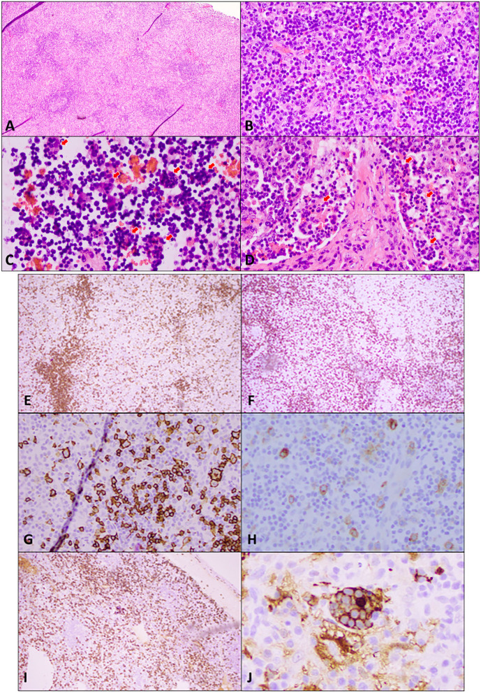
Figure 2.
Excisional biopsy of an enlarged supraclavicular lymph node. (A) Hematoxylin and eosin (H&E) staining at low power (4×) shows widely spaced reactive follicles with a prominent paracortex expansion. (B) H&E medium power (20×) view demonstrates a mixed small and large atypical lymphocytic infiltrate in the paracortex against the background of abundant plasma cells, immunoblasts, and histiocytes. (C) H&E high power (40×) view of cells in sinuses shows numerous histocytes with vacuolated nuclei and abundant cytoplasm engulfing lymphocytes and other cells in a process termed emperipolesis (red arrows). (D) H&E high power (40×) view of the stain demonstrates emperipolesis (red arrows) in sinuses with adjacent areas of fibrosis. Immunohistochemical studies: (E) A low power (10×) view of CD20 immunostaining shows disrupted reactive follicles and scattered large atypical B-lymphocytes. (F) A lower power (10×) view of CD3 immunostaining highlights the admixed paracortical T-cells. (G) A medium power (20×) view of the CD20 immunostaining is positive in atypical lymphocytes. PAX 5 also stains the large atypical cells (not shown). (H) CD30 immunostaining (20× power) highlights the large atypical lymphocytes in interfollicular areas, which were negative for CD15 (not shown). This makes Hodgkin's lymphoma unlikely. (I) CD138 immunostaining (10×) demonstrates the presence of abundant plasma cells, which are also positive for CD79a, MUM1 with polyclonal kappa, or lambda by in situ hybridization (not shown). Plasma cells are often associated with Rosai–Dorfman disease. (J) Areas of emperipolesis are better demonstrated with S100 stain (high power, 60×), which highlights histiocytes with an abundant cytoplasm that have engulfed lymphocytes and other cells (emperipolesis). Engulfed cells do not stain but are outlined by S100 staining when engulfed.

