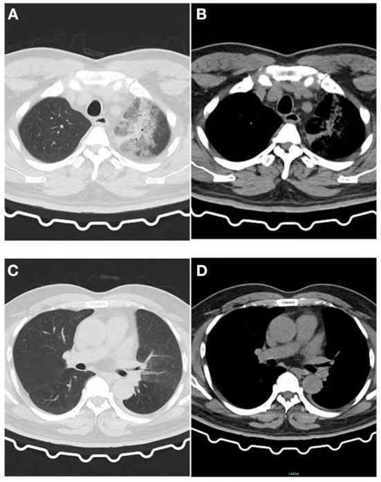
Figure 1.

Image of the patient's first CT inspection on June 5, 2021. (A) Lung window: Patchy high-density shadow in the upper lobe of the left lung with blurred edges. (B) Mediastinal window: Patchy exudative lesions in the upper lobe of the left lung. (C) Lung window: No obvious exudative lesions in the left lower lobe, and a small amount of pleural effusion on the left. (D) Mediastinal window: No obvious exudative lesions.
