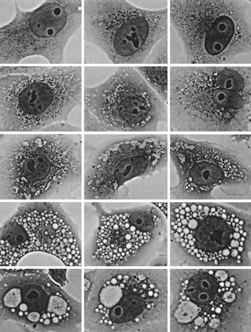
FIG. 1.
Temporal progression of cell vacuolation caused by V. cholerae hemolysin (phase-contrast light microscopy; magnification, ×200). Top row, control cells, not exposed to toxin, showing no vacuolation. Second row, control for the exacerbation of V. cholerae-induced vacuolation by concanamycin (see row 4), showing that only a hint of vacuolation is caused by 1 h of exposure to this proton pump inhibitor alone. Third row, initial, relatively mild vacuolating effect caused by exposure to V. cholerae hemolysin for 1 to 2 h. Characteristically, this involves peripheral elements of the cell that can be identified as components of the ER by separate dye uptake and electron microscopic studies (not shown). Fourth row, more severe, diffuse, bubbly vacuolation caused by simultaneous 2-h exposure to V. cholerae hemolysin (same 1/500 dilution of culture supernatant as in row 3) plus 100 nM concanamycin (same concentration as in row 2). Fifth row, End-stage vacuolation of Vero cells, which appears in some cultures as early as 1 h after exposure to concentrated V. cholerae hemolysin but eventually is produced by all vacuolating strains at longer times of exposure to lower toxin concentrations. The gigantic vacuoles observed in such cells contain vast numbers of very negatively charged internal membranous vesicles, indicating they are multivesicular bodies or autophagosomes.

