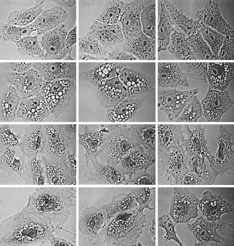
FIG. 4.
The endocytotic tracer protein HRP is denied access to V. cholerae hemolysin-induced vacuoles, and thus they either are not derived from endosomes or are at least “off the circuit” of normal endosomal trafficking. Top row, control cells not exposed to V. cholerae hemolysin. After a 15-min exposure to HRP and a 30-min chase, the tracer has found its way deep into the cells, into the plethora of ∼0.5-μm granules that tend to collect in the perinuclear area, characteristic of normal late endosomes. Second row, cells loaded with HRP exactly like the control cells in the top row, but only after a 60-min exposure to a 1/500 dilution of V. cholerae supernatant. HRP endocytosis has proceeded to a normal extent (as judged by the numbers of dark granules that have formed), but the granules have remained dispersed throughout the cell, suggesting that they have failed to mature into late endosomes. (Parallel time-lapse light microscopic studies show that these endosomes lack the normal microtubule-directed motility that leads to their centripetal migration and maturation.) Note particularly that HRP has not gained access to any of the V. cholerae-induced vacuoles. Third row, cells prevacuolated by exposure to V. cholerae supernatant as in the second row and then exposed to HRP for only 5 min. Various amounts of HRP have entered these cells, from left to right panels, indicating different levels of endocytotic activity. At this early time of HRP entry, endosomes would not have had time to mature and move centripetally, even in normal cells. Hence, they remain scattered among the hemolysin-induced vacuoles but they still appear “unwilling” to fuse with these vacuoles. Bottom row, Controls for the above-described experiments, illustrating (in the left two panels) cells vacuolated with V. cholerae supernatant as described above and then treated for HRP histochemistry without ever exposing them to HRP, to ensure that the unusually large amount of DAB reaction product seen in some cells (such as the right panel of the third row) does not mean that Vero cells are somehow being stimulated by the hemolysin to express endogenous peroxidatic activity. The rightmost panel of this row shows cells exposed to toxin, then exposed to HRP, and then washed for a full 2 h to allow any recovery from vacuolation that might occur. Still, even at this late time point, HRP has not entered the remaining vacuoles.

