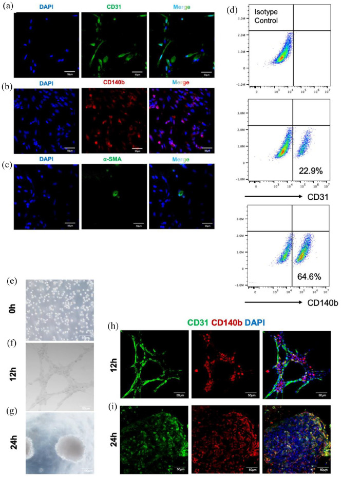
Figure 3.
Identification of VCs from iMVs. (a–c) Immunofluorescence staining indicated ECs (CD31+ in green), pericytes (CD140b+ in red), and vSMCs (α-SMA+ in green) in dissociated iMVs. Scale bar, 50 μm. (d) Flow cytometric analysis for the proportion of vascular cells. CD31 as a surface marker of ECs; CD140b as a surface marker of pericytes. (e–g) Images for tube formation analysis at three different times. After 12 h of seeding, the iMVs-VCs formed a cell-cell network; and 24 h later, the iMVs-VCs self-organized into aggregates. Scale bar, 100 μm. (h) Immunofluorescence of sprouting network showed the interaction between ECs (CD31 in green) and pericytes (CD140b in red) at 12 h. Scale bar, 100 μm. (i) Immunofluorescence of aggregates at 24 h indicated self-organization of ECs (CD31 in green) and pericytes (CD140b in red). Scale bar, 100 μm.

