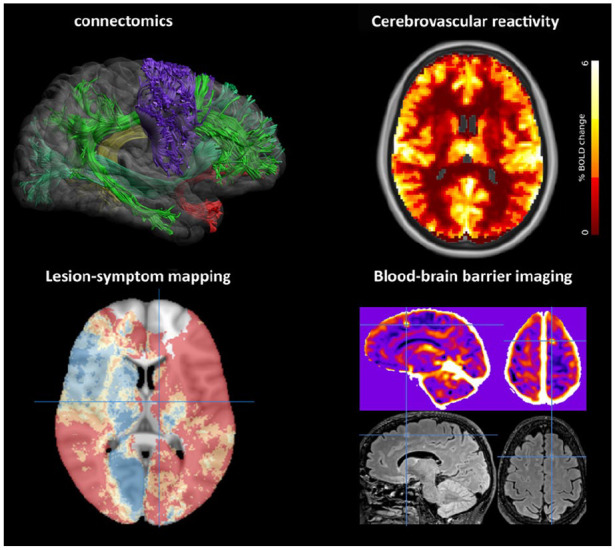
Figure 2.

Emerging brain imaging techniques in VCI. Innovative brain imaging methods to improve detection of functional impact (left side of the figure) and underlying mechanisms (right) of vascular injury in VCI are developing rapidly. Several examples are shown. Connectomics: reconstruction of white matter tracts from diffusion-weighted imaging (image courtesy of Alberto De Luca, UMC Utrecht). Lesion-symptom mapping: brain vulnerability map derived from 2950 ischemic stroke patients, showing the predicted risk (dark blue: lowest risk; red: highest risk) of post-stroke cognitive impairment based on infarct location; crosshair indicates the left thalamus.10 Cerebrovascular reactivity: voxelwise reactivity maps derived from fMRI showing change in Blood-oxygen-level-dependent (BOLD) signal after hypercapnic stimulus (image courtesy of Hilde van den Brink, UMC Utrecht; SVDs@target consortium).32 Blood-brain barrier (BBB) imaging with Dynamic Contrast Enhanced (DCE) MRI (upper panel) showing a frontal BBB leakage hotspot (crosshairs). The corresponding FLAIR image (lower panel) shows a hyperintense lesion at the same site (image courtesy of Michael Thrippleton and Joanna Wardlaw, University of Edinburgh, SVDs@target consortium).
