Figure 5.
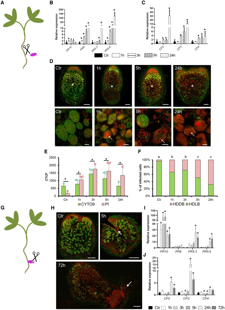
Wounding triggers defense and senescence activation in fix+ nodules associated with the death of the differentiated bacteroids. A, In the first wounding treatment the WT nodules at 21-dpi inoculated with S. medicae WSM419 were separated from the roots. Expression analysis of (B) PR and (C) CP genes after incubation of 0 (Ctr), 1, 3, 5, and 24 h revealed that PRs and CPs are, respectively, induced after 1 and 3 h. D, Observation of bacteroid survival using live (green [SYTO9]) and dead (red [PI]) staining in wounded 21-dpi nodules after 0 (Ctr), 1, 3, 5, and 24 h of incubations reveals a death of the differentiated bacteroids 1 h after incubation which increases with time. Top panel displays the nodule sections (scale bars are 200 µm) and bottom panel shows the bacteroids in the fixation zone III (scale bars are 20 µm). Asterisks indicate the nitrogen-fixation zone and the arrows show dead bacteroids. E, The CTCF of SYTO9 and PI staining calculated from nodule sections of wounded nodules reveals more PI than SYTO9 staining in 1, 3, 5, and 24 h compared to the reference (Ctr). The CTCF were calculated for each time of incubation on five to seven sections of independent nodules and error bars show the se. Asterisks show significant variation between SYTO9 and PI fluorescence and the letters show statistical groups between incubation times using Student’s t test (P < 5%). F, The percentage of nodule infected cells with HDDB or HDLB is calculated in the ZIII of sections from wounded nodules at 0 (Ctr), 1, 3, 5, and 24 h. Augmentation of HDDB cell proportion is observed as early as 1 h and increases during the time of incubation. The proportions of HDDB and HDLB were calculated on the nodule sections used in the CTCF determination. The analysis was performed on five to seven sections collected from nodules of independent plants. The letters show statistical groups between incubation times using Student’s t test (P < 5%). G, The second wounding treatment consists of cutting WT nodules attached to the roots at 21-dpi with S. medicae WSM419. H, Observation of bacteroid survival using live (green) and dead (red) staining in wounded 21-dpi nodules after 0 (Ctr), 1, 3, 5, 24, and 72 h of incubation reveals that bacteroid death starts at 5 h after incubation and is located around the wounded zones. The arrows show the wounded zones and the scale bars represent 250 µm. Expression analysis of the PRs (I) and the CPs (J) shows the upregulation of most of these genes after 24 h of incubation. The expression analysis in B, C, I, and J corresponds to the mean expression of three independent experiments (eight plants per experiment) with two to three technical replicates. The actin housekeeping gene was used for expression normalization. Error bars indicate se and asterisks represent significant variation (P < 2.5%) compared to the WT using the Mann–Whitney statistical test.

