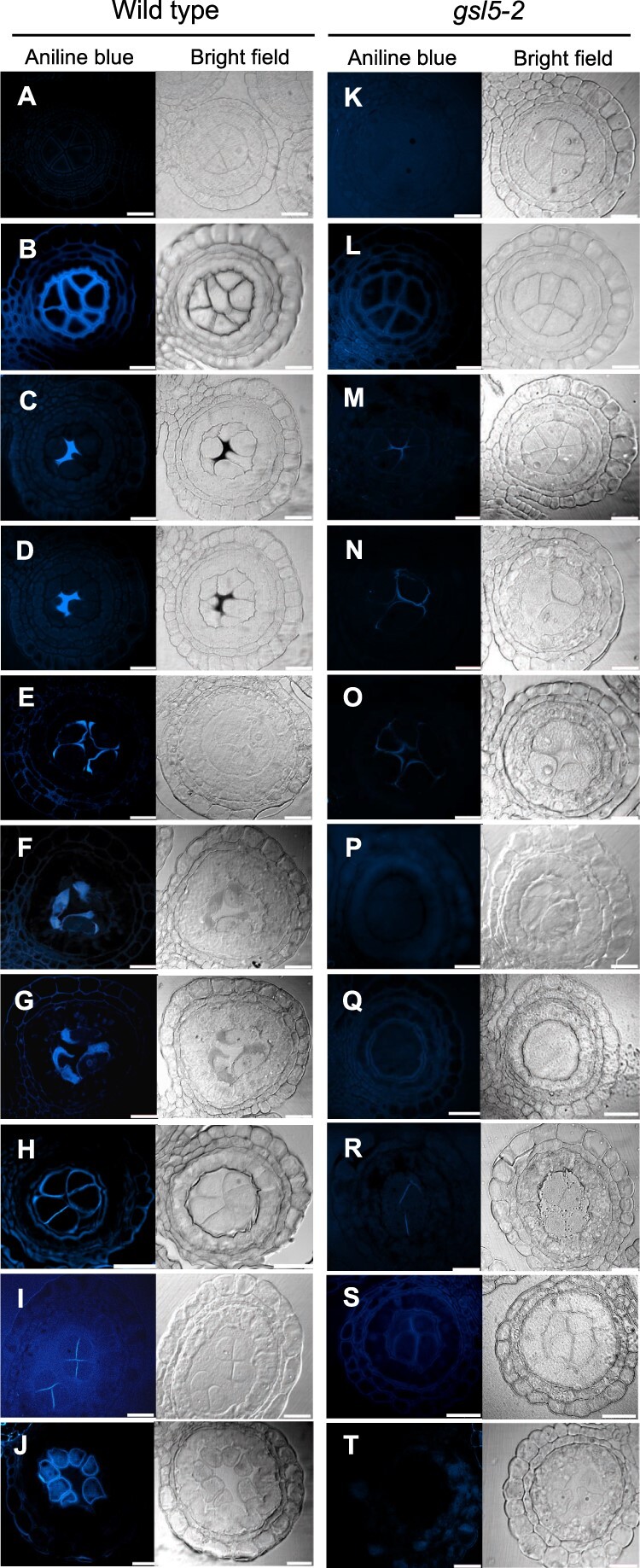
Figure 3.

Callose accumulation during male meiosis in WT and Osgsl5-2 anthers. Callose accumulation pattern was monitored by aniline blue staining at different male meiosis substages. A and K, Mitotic SPC stage; B and L, Premeiotic interphase; C and M, Leptotene; D and N, Zygotene; E and O, Pachytene; F and P, Diplotene; G and Q, Diakinesis; H and R, Dyad; I and S, Tetrad; J and T, Microspore. An arrowhead in the inset in (B) indicates the cell wall unstained with aniline blue. Bar = 20 µm.
