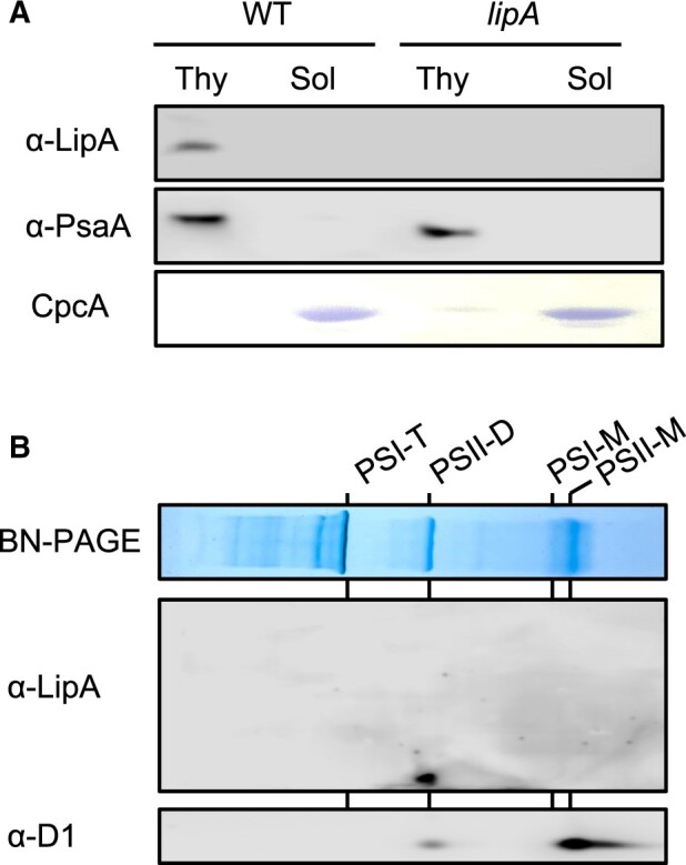
Figure 5.

Localization of LipA protein. A, Distribution of LipA protein in fractions of thylakoid membranes or soluble proteins from WT and lipA cells analyzed with a specific antibody. PsaA and C-phycocyanin alpha chain (CpcA) proteins were used as the controls for thylakoid and soluble proteins, respectively. B, Localization of LipA protein in photosynthetic complexes in thylakoid membranes. Photosynthetic complexes in thylakoid membranes (corresponding to 8 μg chlorophyll) prepared from WT cells exposed to strong light condition for 20 min were separated on BN-PAGE, and the distribution of LipA protein in photosynthetic complexes was analyzed with specific antibodies. PSI-T, PSI trimer; PSII-D, PSII dimer; PSI-M, PSI monomer; and PSII-M, PSII monomer.
