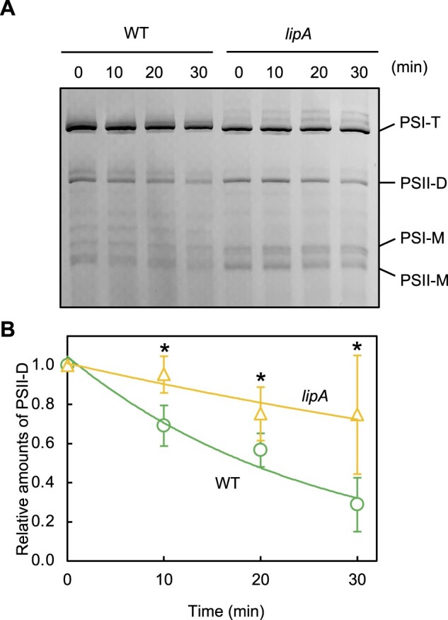
Figure 6.

Roles of LipA in PSII complexes in vivo. A, Photosynthetic complexes in WT and lipA cells analyzed by BN-PAGE. Thylakoid membranes were prepared from WT and lipA cells that were exposed to strong light at 1,500 μmol photons m−2 s−1 (SL) at 32°C in the presence of 200 μg mL−1 lincomycin. Photosynthetic complexes were separated on BN-PAGE. PSI-T, PSI trimer; PSII-D, PSII dimer; PSI-M, PSI monomer; and PSII-M, PSII monomer. B, The relative level of PSII-D was quantified densitometrically. Asterisks indicate statistically significant differences between WT and lipA cells at the point of 30 min (P < 0.01, Student’s t test).
