Figure 3.
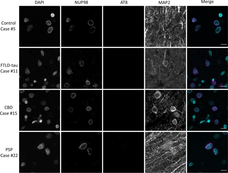
Localization of NUP98 in primary tauopathy occipital cortex. Calcarine cortex (Brodmann area 17) sections from cognitively normal controls, FTLD-tau, CBD, or PSP cases were stained for NUP98, phospho-tau (AT8) and MAP2 by immunofluorescence and nuclei were counterstained with DAPI. Merged images demonstrate DAPI in cyan, NUP98 in magenta and AT8 in green. MAP2 is omitted from the merged images for the sake of clarity. Scale bar = 10 µm.

