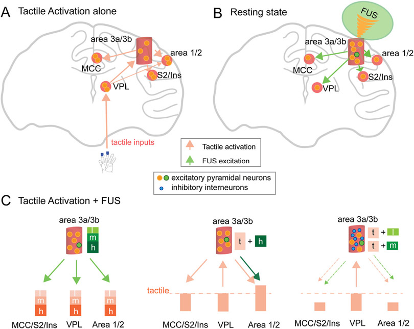
Fig. 7. Schematic model of neuron type selective modulation of 250 kHz FUS.
FUS modulation of two types of neurons is proposed: excitatory pyramidal neurons (large orange and green dots) and inhibitory interneurons (small blue dots). (A) Tactile stimulation of digits elicited peripheral neuron activation that projected to the thalamic VPL nucleus, then to area 3a/3b, area 1/2, and then to MCC and S2/insula. Light orange arrows indicate the direction of information flow between regions. (B) FUS directly activates area 3a/3b neurons and then the activation propagates to off-target regions. (C) Illustrations showing the net outcomes of neural signal changes at three groups of off-target regions: area 1/2, MCC/S2/insula, and VPL, and their connections to area 3a/3b. Arrows indicate the directions of activation propagation. Line thickness indicates the general strength of information flow. The dotted line indicates the magnitude of tactile stimulation evoked activation. Each of the three brain region groups represents different degrees and patterns of feedforward and feedback connections (indicated by colored arrows and directions). (Left) Three intensities of FUS-evoked graded neural activity at the targeted area 3a/3b and off-target regions at resting state. (Middle) In the high-FUS + tactile stimulation condition, high-FUS may predominantly activate a larger proportion of excitatory neurons at area 3a/3b evidenced by the lack of inhibition at this target area. Therefore, tactile responses remained unchanged in all regions except for an enhanced response in area 1/2, which receives extensive feedforward connections from area 3a/3b. (Right) Medium- and low-intensity FUS likely activated more inhibitory than excitatory neurons, therefore reducing the output signals from area 3a/3b (middle). Areas that receive inputs from area 3a/3b via feedforward connections (area 1/2, MCC, S2, and Insula) all exhibited reduced tactile signals. Tactile-evoked neural activation signals at VPL were not affected because it is a signal feeding region to area 3a/3b via thalamocortical afferents. Signal reductions in area 3a/3b has little effect on VPL.

