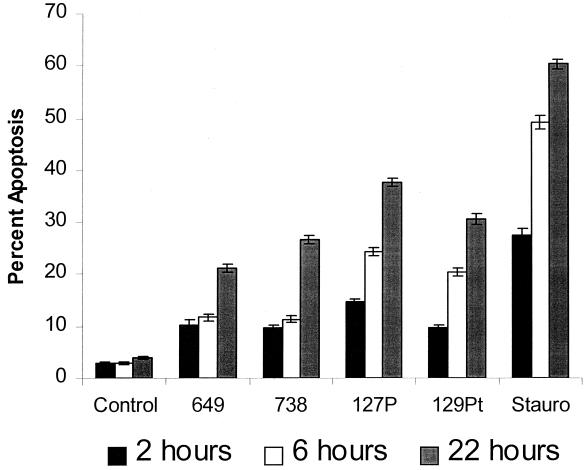
FIG. 8.
H. LOS induces bovine endothelial cell apoptosis. Bovine pulmonary artery endothelial cells (5 × 10⁴) were incubated with 500 ng of LOS/ml isolated from pathogenic isolates (649 and 738) and asymptomatic carrier isolates (127P and 129Pt) of H. somnus for 2, 6, and 22 h at 37°C with 5% CO2. Endothelial cells incubated in medium alone (control) or with staurosporine (200 nM; Stauro) were used as negative and positive controls, respectively. Apoptosis was detected by Hoechst 33342 staining. Five random areas per coverslip were sampled, and 100 random cells were analyzed for apoptosis. The data illustrate the mean ± SEM of three separate experiments. Statistical analysis was performed by ANOVA followed by the Tukey-Kramer significant difference test. Asymptomatic carrier and pathogenic isolate LOS used in these experiments induced significant (P < 0.05) levels of endothelial cell apoptosis at each time point, compared to those of untreated (Control) cells.

