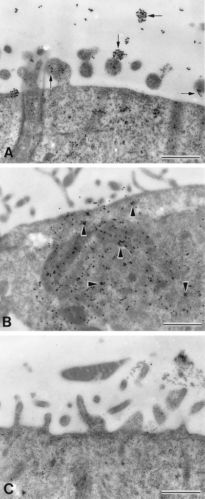
FIG. 3.
Immunoelectron photomicrographs of Caco-2 cells incubated for 2 min (A) or 6 min (B and C) with CSL and then probed with MAb 3E2 (A and B) or isotype-matched control MAb (C). Note dense multifocal immunogold labeling of surface microvilli (arrows) initially (A), followed by progressively increased intracellular labeling (arrowheads) over time (B). Bars, 1 μm.

