
Fig. 3.
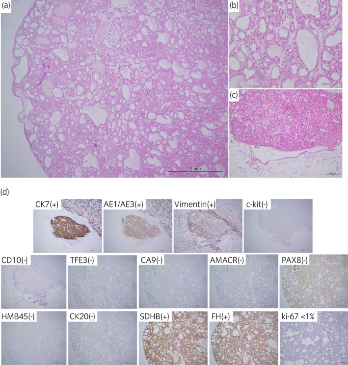
(a) The tumor showed tubular, microcystic growth patterns. (b) Tumor cells had round nuclei and eosinophilic cytoplasm. Perinuclear halo and capillary networks were not prominent (H&E staining). (c) Venous invasion was detected at the fat tissue in the renal pelvis (H&E staining). (d) Tumor cells were positive for CK7, AE1/AE3, and vimentin, and negative for c‐kit, CD10, TFE3, CA9, AMACR, PAX8, HMB45, and CK20. SDHB and FH expression were retained (positive). Ki‐67 labeling index was less than 1%.