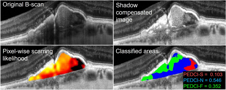
Figure 2.
Classification of pixels within a pigment epithelial detachment (PED) of a patient with neovascular age-related macular degeneration. The original B-scan was taken using spectral domain optical coherence tomography. Results from subsequent shadow compensation and computed per-pixel scarring likelihood are shown. Classified areas include serous (red), neovascular (blue), and fibrous (green) components of the PED. Classified areas were used to compute the PED composition indices: serous (PEDCI-S), neovascular (PEDCI-N), and fibrous (PEDCI-F).

