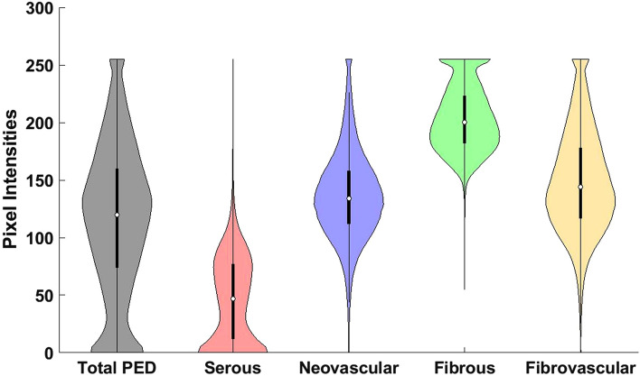
Figure 3.
Violin plot showing the distribution of pixel intensities from different components of a pigment epithelial detachment (PED). Pixel intensities correspond to pre-processed spectral-domain optical coherence tomography images taken from 43 eyes with neovascular age-related macular degeneration. Mutually exclusive classified components include serous, neovascular, and fibrous. Fibrovascular is the combined distribution of neovascular and fibrous tissue. Total PED is the combined distribution of serous, neovascular, and fibrous tissue. The overlap in the distributions indicates the difficulty in separating tissue types, particularly neovascular and fibrous tissue. The skewed distribution of fibrovascular tissue indicates more than one underlying tissue type.

