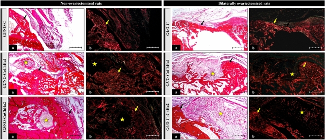
Figure 4.
Photomicrographs of histological slides stained with Picrosirius red obtained from the groups of non-ovariectomized and bilaterally ovariectomized rats and examined under an optical microscope (a) and under polarized light (b) at ×10 magnification. The black arrows point to areas of bone neoformation. The yellow arrows indicate the presence of collagen fibers in the extracellular matrix. The star indicates the area occupied by remnant biomaterial after bioresorption. Scale bar: 20 µm.

