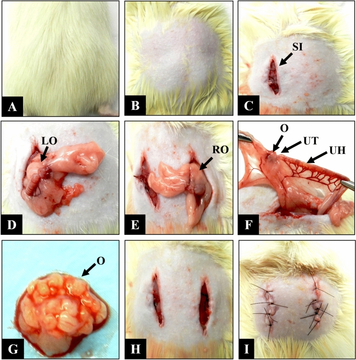
Figure 6.
Ovariectomy of female Wistar rats. (A) Positioning of the animal in ventral decubitus. (B) Shaving of the lumbar region. (C) Longitudinal 10-mm surgical incision on the left side (SI). (D) Exploration of the left ovary (LO). (E) Contralateral posterior incision and exploration of the right ovary (RO). (F) Identification of the ovary (O), uterine tube (UT), and uterine horn (UH). (G) Dissection of the ovary (O). (H) Repositioning of the anatomical structures. (I) Suture of muscles and skin, respectively.

