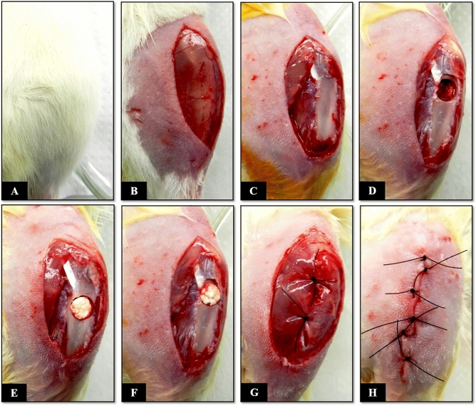
Figure 7.
Surgical procedure for creation of a proximal metaphyseal bone defect in the tibia of Wistar rats. (A) Positioning of the rat in dorsal decubitus, with the left knee flexed. (B) Shaving of the left leg, followed by a longitudinal 15-mm skin incision. (C) Longitudinal incision in the muscle and periosteum. (D) Creation of a 2.5-mm bone defect in the proximal metaphysis of the left tibia. (E and F) Grafting with the CoChHa1 and CoChHa2 scaffolds. (G) Repositioning of the periosteum and muscular suture. (H) Skin suture.

