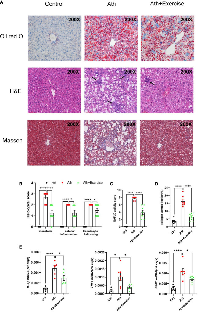
Figure 2.
Aerobic exercise decreases hepatosteatosis, lobular inflammation and fibrosis in mouse livers. (A) Representative images of liver sections stained by Oil Red O, HE and Masson staining from control, NASH and NASH+Exercise mice (original magnification, ×200). (B) Histological scores of steatoses, lobular inflammation, and hepatocyte ballooning in H&E-stained livers. (C) NAFLD activity score was calculated by the histological score of steatosis, lobular inflammation, and hepatocyte ballooning. (D) The collagen volume fraction by Masson staining. (E) Relative mRNA levels of IL-1β, TNFα and F4/80 in liver tissues from control, NASH and NASH+Exercise mice. Data are depicted in mean ± SEM. *P<0.05; ****P<0.0001.

