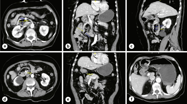
Fig. 1.
CT abdomen pelvis demonstrating a 2.7 × 3.7 × 5.2 cm complex pancreatic head lesion containing air and debris (a), contiguous with the duodenum at the junction of the second and third segments (b) with moderate 1.0 cm dilatation of the common bile duct (c) and 0.6 cm dilatation of the main pancreatic duct (d, e). CT abdomen pelvis demonstrating GOO manifested as markedly distended stomach with distal gastric and proximal duodenal wall thickening and hyperenhancement (f).

