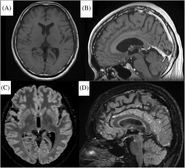
FIGURE 1.

Cerebral magnetic resonance images obtained on the third day: (A) and (B) Axial and sagittal images of the Gd‐enhanced T1‐weighted sequence show no abnormal findings. (C) and (D) Axial and sagittal images with the Gd‐enhanced Cube FLAIR sequence show nodular lesions with high intensity scattered on the meninges of the frontal and occipital lobes
