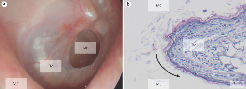
Fig. 1.
Epithelialization of TMPs. a Ear microscopy of a TMP shows round epithelialized margins in the TMP. b Histology (H&E) of a TMP confirms epithelialization of the defect. The black arrow illustrates the overgrowth of the keratinizing squamous epithelium over the defect margin. EAC, external auditory canal; ME, middle ear; TM, tympanic membrane; TMP, tympanic membrane perforation.

