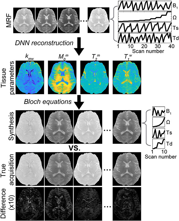
Figure 13. Synthetic MRI analysis for validation of the semisolid MT-MRF method.
Synthetic contrast-weighted images are generated using all the tissue parameters obtained from the deep neural network (DNN), which can then be compared with the experimentally acquired images as the standard of reference. Tissue parameters are quantified from an acquisition schedule consisting of 40 dynamic MRF images (a corresponding MRF schedule is shown in top right) using the DNN, and then a new acquisition schedule (middle right) is used for synthesizing 10 dynamic MRF images by inserting the tissue parameters obtained from DNN into the forward BM transform. The synthesized images showed a high degree of agreement with the experimentally acquired images as shown in the difference image. Reproduced and modified with permission from Kim et al., Neuroimage 2020:117165.73

