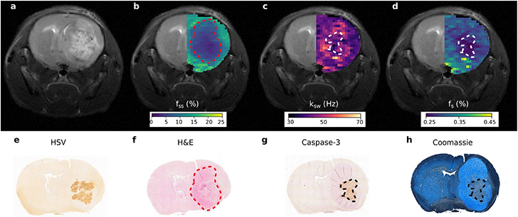
Figure 7. Quantitative imaging of apoptosis following oncolytic virotherapy using sequential and deep semisolid MT/CEST MRF.
a. Conventional T2-weighted image of an oncolytic virotherapy treated mouse, 72 hours post virus inoculation, is incapable of detecting treatment responsive apoptotic regions. b. Semisolid macro-molecules proton volume fraction (fss) map, where a decreased volume-fraction represents tumor-related edema and a change in the lipid composition of tumor tissue relative to normal brain tissue. c. Amide proton exchange-rate (ksw) and d. volume fraction (fs) maps. Regions of decreased intracellular pH and mobile protein concentration, respectively, are indicative of apoptosis. e-h. Histology and immunohistochemistry images validate the MR findings with cleaved caspase-3 positive tumor regions and decreased Coomassie blue protein staining, indicative of apoptosis, colocalizing with the regions of decreased exchange rate and mobile protein concentration. Reproduced with permission from Perlman et al. Nat Biomed Eng. 2021;1-10. https://doi.org/10.1038/s41551-021-00809-7.15

