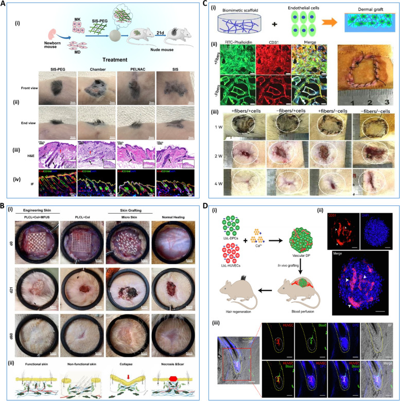
Fig. 5.
3D bioprinting for trauma repair and hair follicle regeneration A SIS-PEG promotes skin wound healing and successful hair growth in mice [179]. (i) schematic diagram of the mechanism, (ii) macroscopic schematic diagram of skin hairs in mice at 21 days, (iii) H&E staining of regenerated skin, (iv) immunofluorescence staining of regenerated skin. B PLCL + COL + MFUS (a tissue engineering functional skin by carrying MFUS in 3D-printed polylactide-co-caprolactone scaffold and COL gel) promotes healing of full-thickness skin defect wounds [180]. (i) healing of full-thickness skin wounds in four groups of mice with PLCL + COL + MFUS, PLCL + COL, micro-skin and conventional treatment on days 0, 21 and 60, (ii) schematic diagram of the mechanism of wound healing in each group. C macroporous filamentous protein scaffold with nanofibrous microstructures promote neovascularization and dermal reconstruction [182].(i) Schematic diagram of in vivo experiments of endothelial cells-seeded nanofibrous scaffolds, (ii) growth and distribution of seeded cells on the scaffolds, (iii) macroscopic observation of wounds in SD rats at the first, second and fourth weeks after scaffold implantation. D layer-by-layer DP spheroids are able to form good blood perfusion in vivo [183] (i) schematic diagram of the mechanism by which layer-by-layer DP spheroids are vascularized in vitro and blood perfusion is formed in vivo, (ii) immunofluorescence showing angiogenesis after 3 days of in vitro culture of layer-by-layer DP, and (iii) immunofluorescence showing blood perfusion after three weeks of in vivo transplantation of layer-by-layer DP. Reprinted with permission from Ref. [179, 180, 182, 183]

