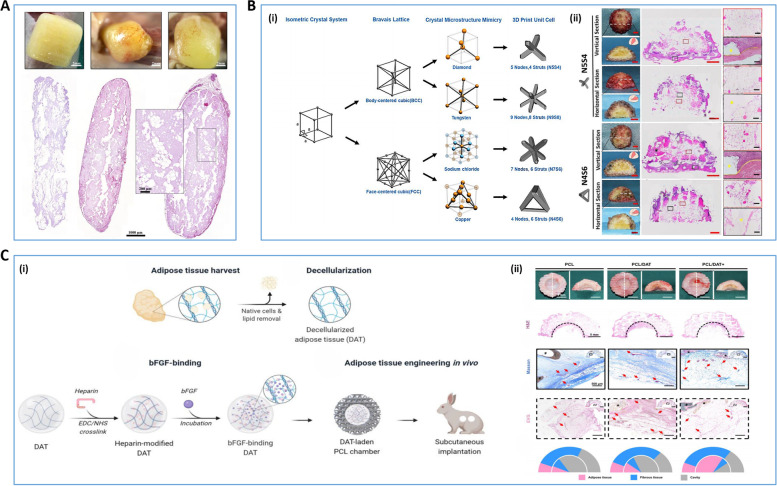
Fig. 8.
3D bioprinting for Breast Implants A Histological images and macroscopic images of freshly printed 3D bioprinting LAT products and 3D bioprinting LAT products cultured in vivo for 30 days and 150 days [202]. B The survival rate of fat in different breast scaffolds was different in nude mouse model experiments [203]. (i) Schematic design of crystal microstructure of unit cells. (ii) After 12 weeks of fat grafting, comparing macroscopic images and H&E staining images of N5S4 and N4S6 groups, the adipose tissue in the N5S4 group had a more regular shape and better integration of the scaffold with adjacent tissues, with less compression. C The Macroporous chambers facilitate large volume soft tissue regeneration from adipose-derived extracellular matrix [204]. (i) Mechanisms to promote the generation of large soft tissue volumes in the ECM of adipocytes using large pore chambers, (ii) Morphological performance of grafted specimens from the PCL, PCL/DAT, and PCL/DAT + groups after 12 weeks, with PCL/DAT + showing better vascularization and more adipose tissue generation. Reprinted with permission from Ref. [202–204]

