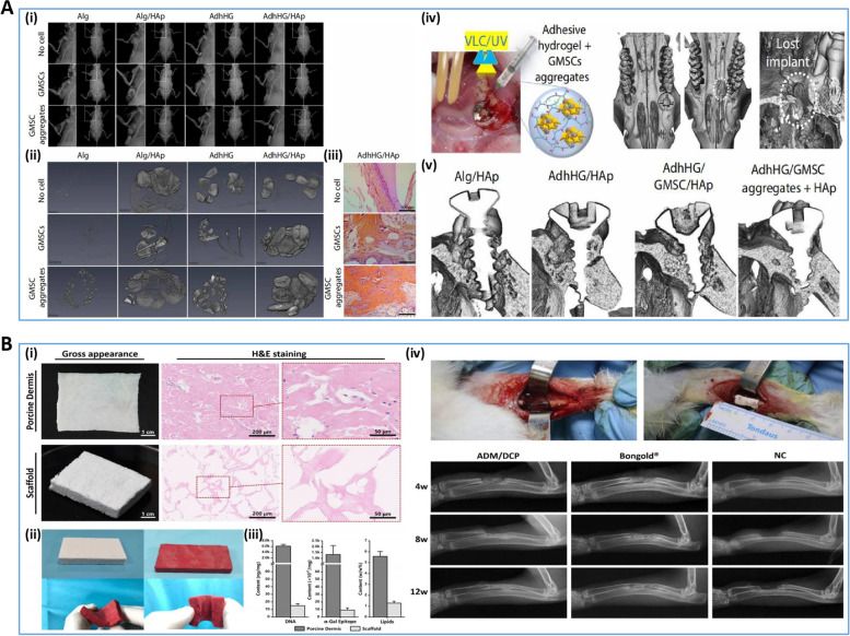
Fig. 9.
3D bioprinting for Maxillofacial Bone Restoration. A Experimental demonstration of bone regeneration capacity of Alg-based adhesive hydrogel (AdhHG)/ Gingival MSCs (GMSC) aggregates + hydroxyapatite (HAp) [213]. (i) Four groups of Alg, Alg/HAp, AdhHG and AdhHG/HAp were mixed with cell-free formulations, GMSC and GMSC aggregates to prepare different hydrogels for implantation under the skin, respectively, and 2D radiographs were observed, (ii) 3D reconstructed images of each group of CT imaging, (iii) H&E staining pictures of each group after 8 weeks of subcutaneous implantation of hydrogels mixed with GMSC, GMSC aggregates, or cell-free formulations of AdhHG/HAp, (iv) Actinomycetes-coated titanium implants cause peripheral inflammation and defects, hydrogels mixed with different formulations are injected into oral defects, (v) 8 weeks after implantation of hydrogels, showing that hydrogels containing different formulations promote regeneration [213]. B Performance demonstration of 3D porous scaffolds based on dicalcium phosphate decellularized dermal matrix [215]. (i) macroscopic appearance and H&E staining of porcine dermis and prepared scaffold to reflect their nucleus-free components, (ii) blood absorption performance of scaffold, (iii) DNA, a-Gal epitope and lipid content display of porcine dermis and scaffold, (iv) X-ray schematic diagram of the middle bone defect of the middle radius in the Dermis-derived matrix/dicalcium phosphate, and negative control group at the 4th, 8th and 12th weeks after surgery, to compare the ability to promote bone regeneration. Reprinted with permission from Ref. [213, 215]

