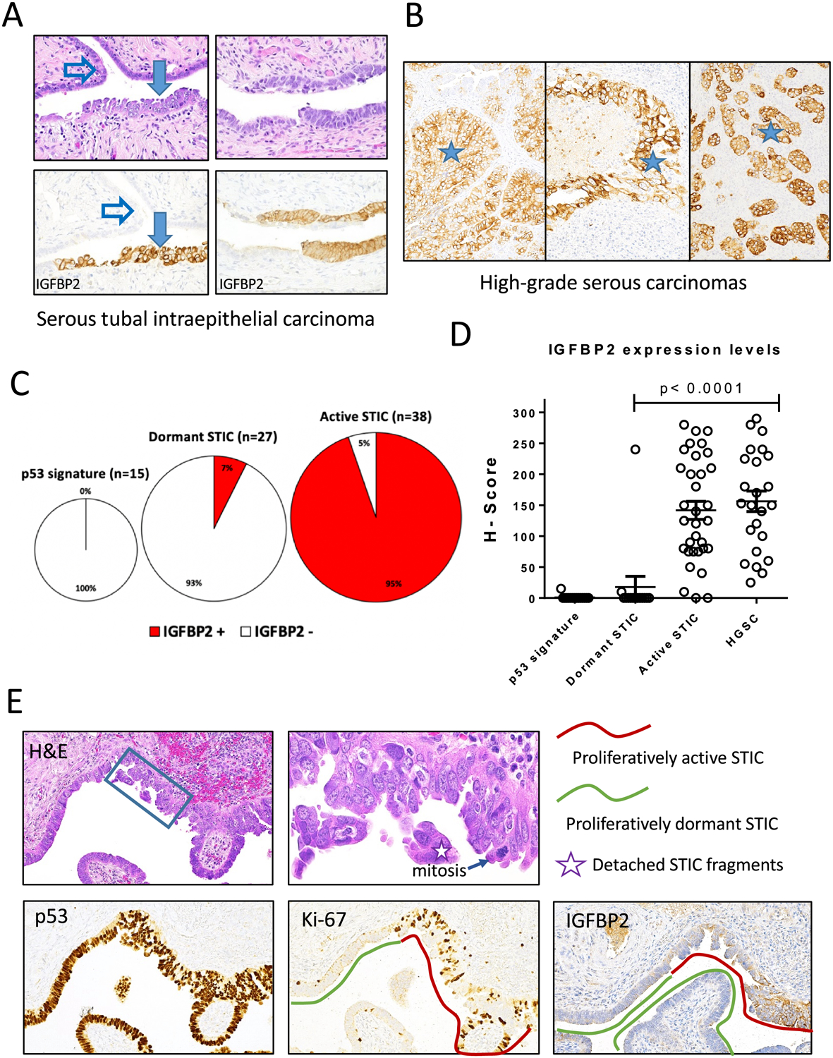
Figure 4. Immunohistochemistry of IGFBP2 and correlation with DNA methylation.

(A-B) Examples showing IGFBP2 immunostaining in STIC (A) and HGSC (B) but not adjacent normal fallopian tube epithelium (NFTE). Solid arrow, STIC; open arrow, NFTE; asterisks, high-grade serous carcinomas in three different cases. (C) Summary of IGFBP2 staining in p53 signature, dormant STIC, and active STIC. Number and percentage of positive cases are shown. (D) Scatter plot of IGFBP2 immunostaining H-scores of various lesions. Each symbol represents an individual lesion. (E) An example of STIC containing both proliferatively active and dormant areas. IGFBP2 immunostaining is positive in proliferatively active STIC (Ki-67 positive).
