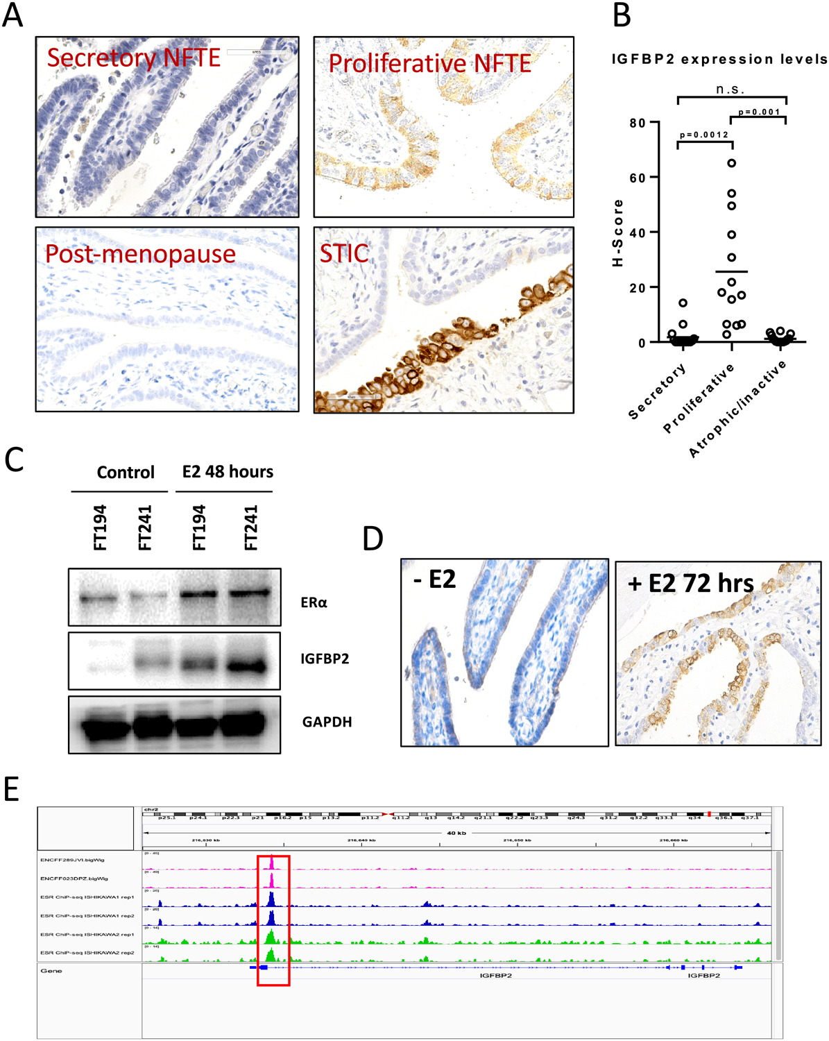
Figure 5. Estrogen regulates IGFBP2 expression in normal premenopausal fallopian tube epithelium.

(A) IGFBP2 immunoreactivity in fallopian tubes from women at different menstrual phases (secretory/proliferative/postmenopausal) and in STIC. (B) H-score of IGFBP2 expression levels at different menstrual phases. (C) Immunoblots for ER-alpha and IGFBP2 in cellular extracts of FT194 and FT241 cell lines after treatment with 50 nM estradiol (E2) for 48 hours. GAPDH is used as a loading control. (D) IGFBP2 expression increases in freshly harvested human fallopian tube tissue after E2 treatment. (E) Chromatin immunoprecipitation-sequencing data for ER-α on T47D and Ishikawa cell lines showing ESR1 peaks (red squares) located within 1 k.b. downstream of the TSS of IGFBP2. The data were obtained from the ENCODE database.
