
Fig. 2.
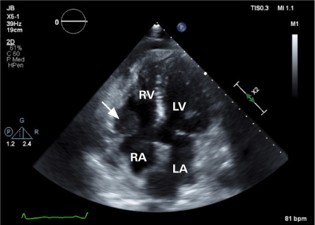
Transthoracic echocardiogram before chemotherapy. A 2.5- × 1.8-cm fixed, hypodense mass (arrow) is attached at the base of the right ventricle.
LA, left atrium; LV, left ventricle; RA, right atrium; RV, right ventricle.
Supplemental motion image is available for Figure 2.