Fig. 4.

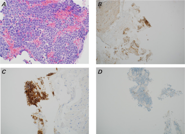
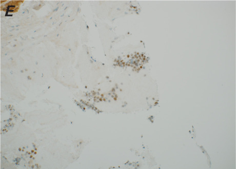
Pathologic samples of the right ventricular mass. A) Hematoxylin and eosin stain show monomorphic lymphocytes with apoptotic bodies that form a “starry sky” appearance that was B) CD10 +, C) CD20 +, D) BCL-2 negative, and E) BCL-6 equivocal. Panel A is at ×40 magnification, and panels B through E are at ×20 magnification. These findings are consistent with Burkitt lymphoma.
Fig. 4.

Pathologic samples of the right ventricular mass. A) Hematoxylin and eosin stain show monomorphic lymphocytes with apoptotic bodies that form a “starry sky” appearance that was B) CD10 +, C) CD20 +, D) BCL-2 negative, and E) BCL-6 equivocal. Panel A is at ×40 magnification, and panels B through E are at ×20 magnification. These findings are consistent with Burkitt lymphoma.
