
Figure 2. Tumor control required CD8+ T cell-intrinsic IRF2 expression.
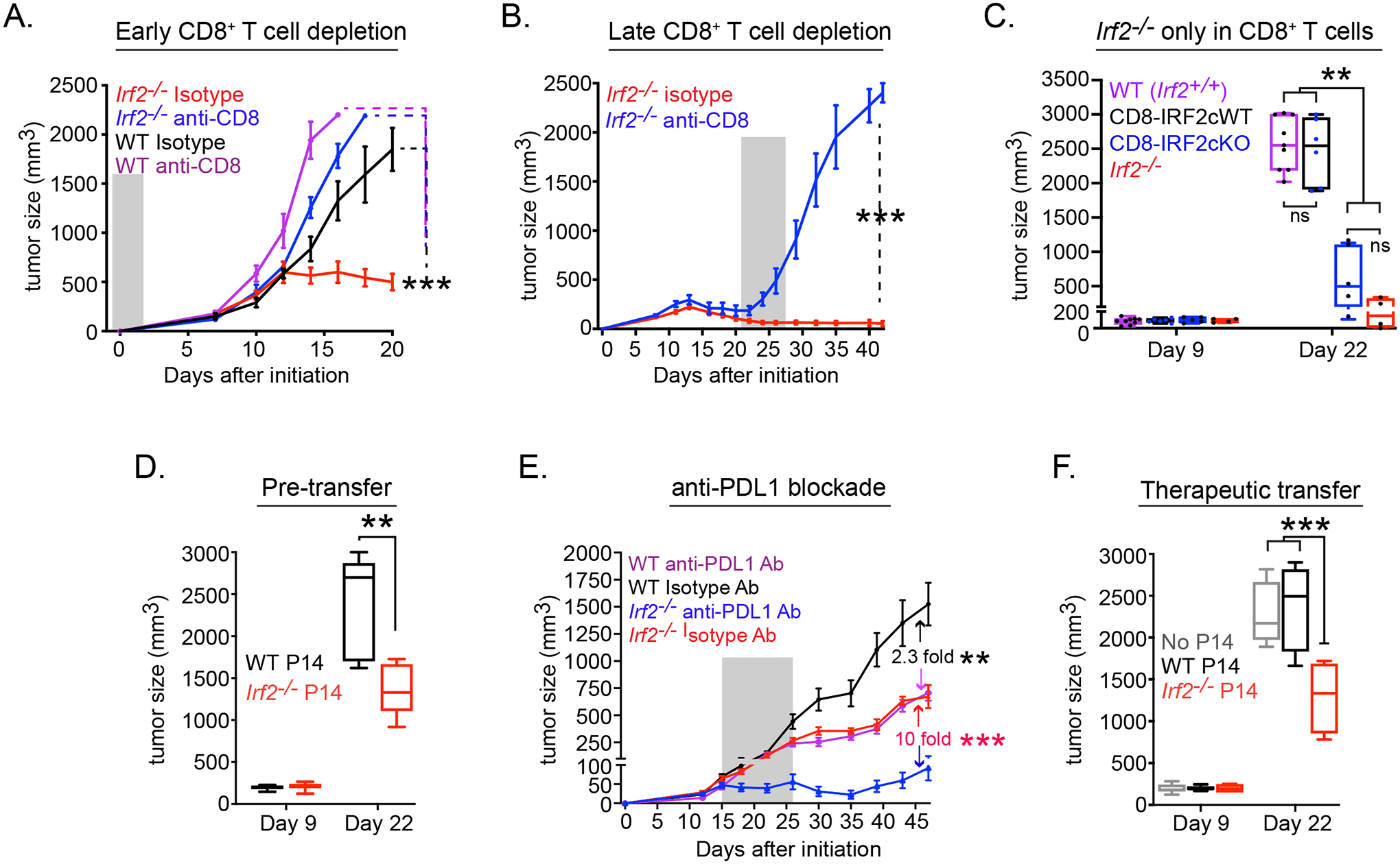
Tumor growth kinetics in WT or Irf2−/− mice that received isotype control or anti-CD8 depleting antibody either (A) one day before (early CD8+ T cell depletion) or (B) 21 days after (late CD8+ T cell depletion) MC38 initiation. For late depletion, only Irf2−/− mice were used since WT mice had already reached endpoint by day 21. Shaded region indicates duration of antibody treatment.
(C) MC38 tumor growth in WT control (i.e., Irf2+/+, purple), CD8-IRF2cWT (i.e., Irf2+/+ CD8Cre+ mice, black), CD8-IRF2cKO (IRF2-deficient only in CD8+ T cells; blue), or Irf2−/− (red) mice.
(D) Tumor size after WT mice received 2×10⁵ naïve WT (black) or Irf2−/− (red) P14 T cells one day prior to receiving MC38-GP tumor.
(E) Tumor growth in WT or Irf2−/− mice with orthotopic PyMT breast tumor cells that were treated with isotype or anti-PDL1 blocking antibody beginning on day 15 after tumor implantation. Number in graph indicates the fold change in tumor size between the isotype vs. anti-PDL1 treatment for WT or Irf2−/− mice. Shaded region indicates duration of antibody treatment.
(F) MC38-GP tumoresize after WT mice received 2×10⁵ pre-activated WT (black) or Irf2−/− (red) P14 T cells (i.v.) on day 9 after tumor initiation.
Data are representative of at least two independent experiments. Error bars represent mean ± SEM. ** p<0.001, *** p< 0.0001. One-way ANOVA for multiple comparisons used for tumor growth kinetics.