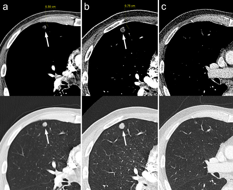
Figure 2.
Axial CT images on soft-tissue and lung window settings demonstrating one of multiple low-density pulmonary nodules within the right lung on initial arterial-phase contrast-enhanced CT (A), which increases in size on a subsequent CTPA (b) and then can be seen to have almost completely resolved following chemotherapy treatment on subsequent restaging contrast-enhanced CT (C). CTPA, CT pulmonary angiography.

