
Figure 2.
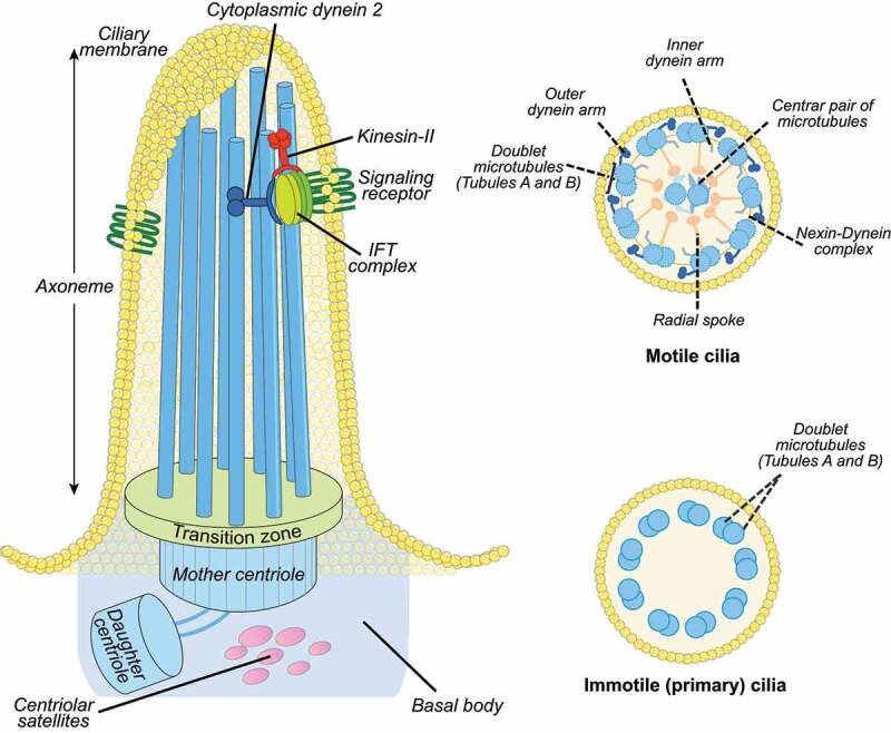
Cilia structure. Left: the ciliary axoneme is composed by nine microtubule doublets of microtubules (blue rods), which nucleate from the basal body (mother centriole). The axoneme protrudes from the cell membrane (ciliary membrane). The movement of IFT particles to the tip of the flagellum is powered by kinesin-II, a microtubule-based molecular motor. The movement of IFT particles back to the base of the flagellum is driven by the cytoplasmic dynein 2, another microtubule-based molecular motor. The mother and daughter centrioles are indicated by the blue cylinders and the transition zone by green at the bottom of the axoneme. The transition zone mediates interactions with the ciliary membrane. Right, top: motile cilia display a central microtubule pair connected with the outer microtubular doublets by radial spokes. The outer microtubules are connected by the nexin-dynein complex. Other structures include the axonemal inner and outer dynein arms. Right, bottom: Immotile primary cilia display nine peripheral microtubular doublets with no central pair and without dynein arms.