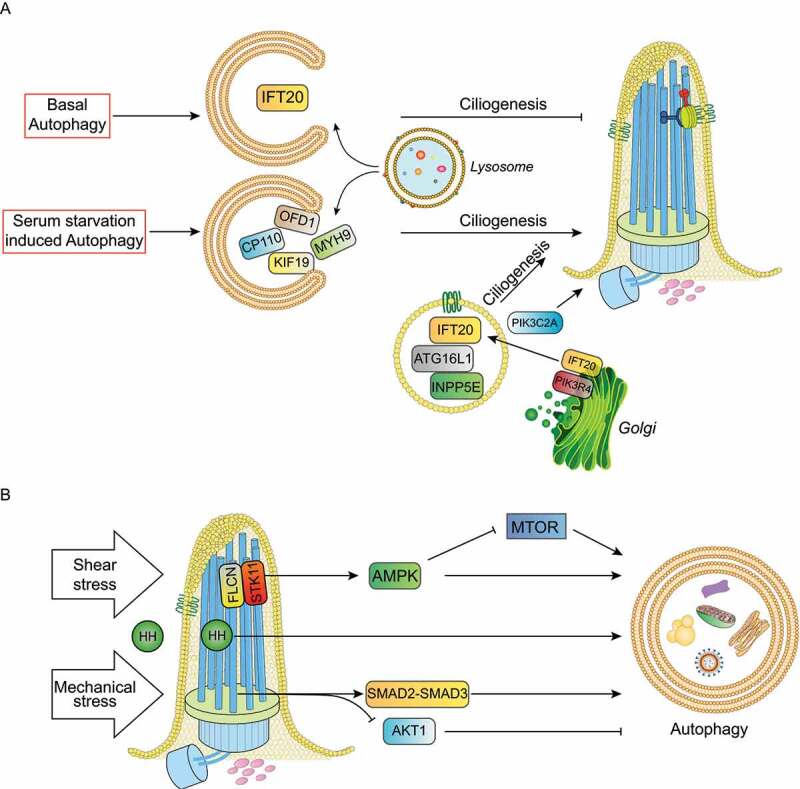
Figure 3.

Cilia-autophagy crosstalk models. (a) Under serum starvation selective autophagy degrades ciliary proteins such as OFD1, MYH9, CP110 and KIF19 to promote ciliogenesis and cilia elongation. Conversely, basal autophagy inhibits ciliogenesis by removing IFT20. ATG16L1, INPP5E and IFT20 trafficking to cilia affects primary cilia organization and signaling independently of their functions in autophagy. PIK3C2A and ATG16L1 control ciliogenesis via PtdIns3P. (b) HH (hedgehog) ligands activate HH signaling transduction and autophagy; shear stress in kidney epithelial cells activate the FLCN-STK11-AMPK-MTORC1 cascade and the mechanical stress activates the autophagy stimulatory SMAD2-SMAD3 pathway and the inhibitory AKT1 signaling in trabecular meshwork cells.
