Figure 3.

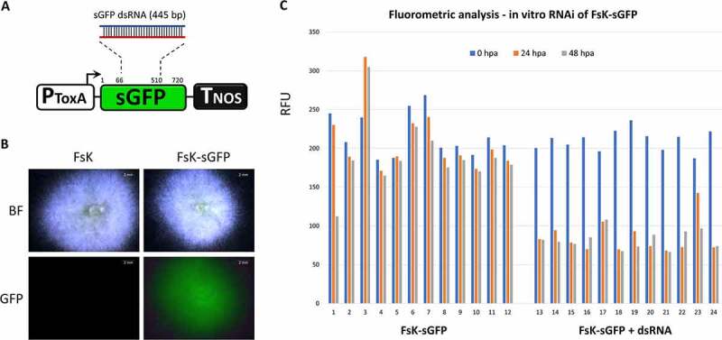
In vitro RNAi in FsK-sGFP. (A) Schematic representation of the sGFP transgene that is present in FsK-sGFP. PToxA: promoter for the promoter from Pyrenophora tritici-repentis ToxA gene; sGFP: GFP variant that contains a serine-to-threonine substitution at amino acid 65; TNOS: terminator for the nopaline synthase gene. The 445 bp fragment chosen for in vitro transcription of dsRNA is depicted. (B) Stereoscopic observation of sGFP fluorescence. (C) Fluorometric analysis for in vitro RNAi in FsK-sGFP. Vertical axis: RFU: relative fluorescence unit, calculated as the ratio of sGFP-indicative fluorescence (excitation 488 nm, emission 515 nm) to growth-indicative absorbance (wavelength 595 nm). Horizontal axis: 1–12: 12 wells containing FsK-sGFP conidia. 13–24: 12 wells containing FsK-sGFP conidia plus 100 ng (each well) sGFP dsRNA.
