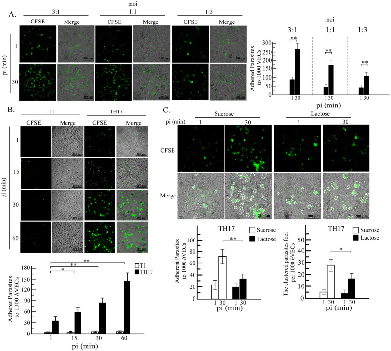
Fig 2. The kinetics of T. vaginalis trophozoites binding to hVECs.
The trophozoites were labeled with CFSE and co-cultured with hVECs. A. trophozoites at a moi of 3 to 1, 1 to 1 or 1 to 3 were incubated with hVECs for 30 min. B. T1 or TH17 trophozoite were co-incubated with hVECs (moi 1:3) for 1, 15, 30 or 60 min. C. trophozoites were co-incubated with hVECs (moi 1:3) in the presence of 250 mM sucrose or lactose for 30 min and after the removal of unbound trophozoites, cell cultures were fixed for imaging by fluorescence and phase-contrast microscopy. PI, post-infection. The clustered foci with over three trophozoites are circled by white-dashed lines. The experiments were performed in triplicate and the average number of binding trophozoite or clustered foci per 1,000 hVECs were measured as shown in the bar graphs (mean ± SD). The statistical difference was analyzed by the Student’s t-test with P<0.05 (*), P< 0.01(**), and ns, not significant.

