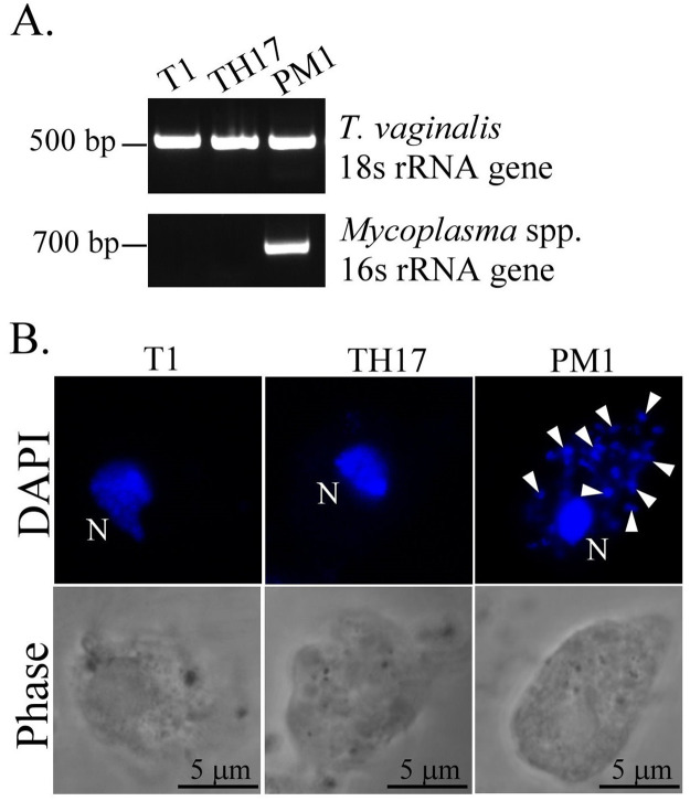
Fig 9. Mycoplasma symbiosis detection in the TH17 isolate.
A. DNA extracted from T1, TH17, and PM1 isolates were amplified by PCR using specific primers for T. vaginalis 18s rRNA or Mycoplasma spp. 16s rRNA genes. The PCR products were separated in a 1% agarose gel. B. The trophozoites from T1, TH17, and PM1 isolates were fixed on a glass slide and stained with DAPI for confocal microscopy. The cell morphology was visualized by phase-contrast microscopy. The T. vaginalis nuclei are indicated by the letter N, and the Mycoplasma DNA puncta are indicated by white arrowheads. Scale bar represents 5 μm.

