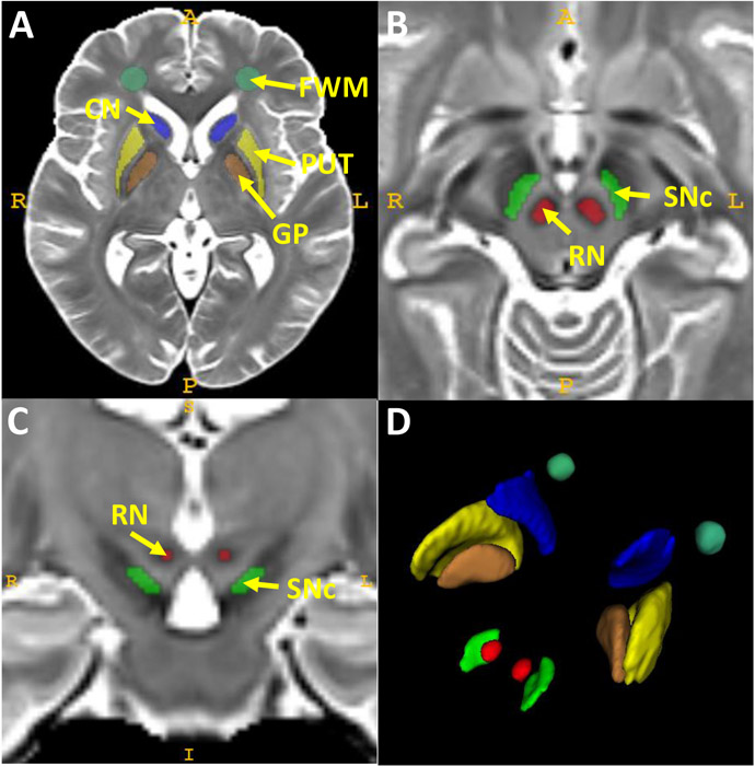
Figure 1. Regions of interest (ROIs) defined on the T2-weighted cohort template.
The locations of the CN, PUT, GP, and FWM are shown in A in an axial slice that captures all four structures. The SNc and RN regions are displayed in B (axial slice) and C (coronal slice) at the midbrain level. A 3D view of all ROIs is shown in D.
Abbreviations: CN: caudate nucleus; FWM: frontal white matter; GP: globus pallidus; PUT: putamen; RN: red nucleus; SN: substantia nigra.

