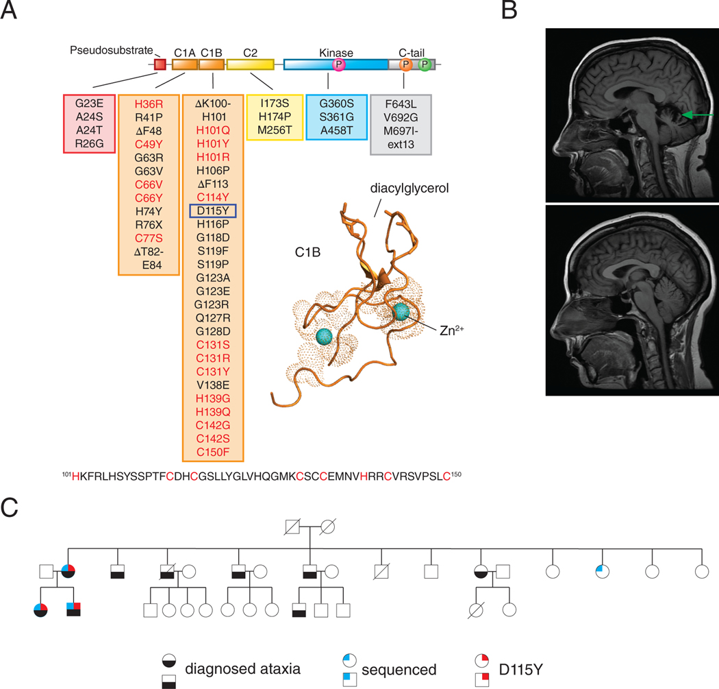
Figure 1. PKCγ in Spinocerebellar Ataxia Type 14.
(A) Primary structure of PKCγ with all known SCA14 variants indicated in boxes beneath each domain (24–27). Newly identified patient variant D115Y is indicated by the blue box. Previously published crystal structure (61) of PKCβII C1B domain is shown with Zn2+ (cyan spheres) and diacylglycerol binding sites labeled (PDB: 3PFQ). Conserved His and Cys residues of Zn2+ finger motif are shown in red in PKCγ primary sequence. (B) MRI of patient at age 46 with D115Y variant (top) compared to age-matched healthy control (bottom); green arrow indicates cerebellar atrophy. (C) Pedigree of family with PKCγ D115Y variant; black shape-fill indicates family members diagnosed with ataxia, blue shape-fill indicate family members that have been sequenced, red shape-fill indicates family members with D115Y variant.

